Box 19162
Arlington, TX 76019-0162
Making (Brain) Waves
The human brain is a mystery. Within an intricate maze of grooves and folds, neurons send and receive billions of signals responsible for vision, touch, hearing, balance, movement, emotion. Stretched flat, the brain would be the size of a small tablecloth.
Our brains serve as a command center for the body. When the brain falters, it can cause dementia, epilepsy, depression, multiple sclerosis, or stroke. Worldwide, some 3 billion people—roughly one in three of us—live with a neurological condition.
Clues to understanding those conditions lay in the brain’s labyrinth. Unraveling how the brain is wired could result in life-changing tools and new approaches to address even the most complex disorders.
That’s why the leaders at The University of Texas at Arlington chose to target brain health for phase one of the Recruiting Innovative Scholars for Excellence, or RISE 100, initiative. Through this multiyear effort, the University will hire experts in the field to complement and bolster the ongoing and groundbreaking brain health research already taking place on campus.
Said research is expansive: Among other projects, UTA faculty are employing light therapy to improve cognition in people at risk for dementia disorders, developing a map of the brain to better understand the progression of Alzheimer’s disease, pinpointing the brain cells that cause epilepsy in children, and studying how to predict if infants could experience a devastating type of brain damage.
New and current faculty now have even more tools at their disposal, as the University opened a $6.2 million Clinical Imaging Research Center in November with a state-of-the-art MRI machine capable of capturing exceptionally detailed images of the brain and other organs.
“It’s hard to overstate the importance of brain health research. Advancements in this field have a direct impact on improving humanity and easing suffering,” says Kate C. Miller, vice president for research and innovation. “By making these investments, we believe UTA will be a beacon for brain health research.”
Strengthening Memory
As we age, our memory often declines. We forget the name of a neighbor or the task that sent us to the kitchen. This is particularly true for working memory, which allows us to maintain multiple facts in our minds at once, falling somewhere between short-term and long-term memory. Working memory is tied to intelligence, concentration, and problem solving. Simply put, working memory is vital to our ability to function day to day.
Hanli Liu, professor of bioengineering, recently found that a noninvasive light therapy called transcranial photobiomodulation (tPBM) can stimulate and increase the power of neurons—and thus that it is capable of strengthening cognitive functions, especially for individuals at risk for Alzheimer’s disease and related dementia disorders.
What makes this discovery so promising is tPBM is low-cost and easy to administer at home.
“As our population ages, healthy aging is more critical than ever,” says Dr. Liu, who also holds a College of Engineering Board of Advisors Endowed Professorship. “Light therapy has the potential to help people live healthy longer.”
“This is a low-cost, non-invasive tool with incredible promise. There’s a real possibility to help people, and maybe even relieve or reduce the progression of dementia disorders.”
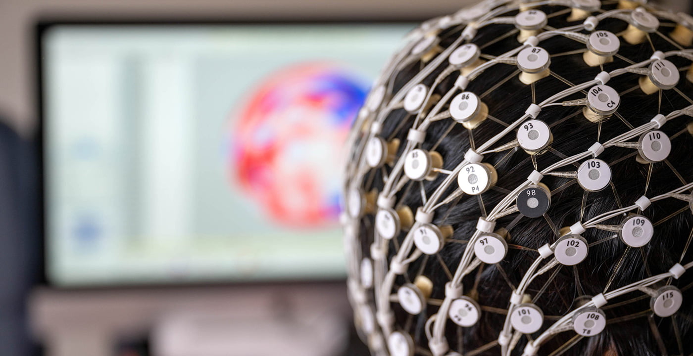
Her study—conducted with an international team of researchers from UTA, Beijing Normal University, and the University of Birmingham—is the first to confirm a neural or neurophysiological link between tPBM and working memory capacity in humans. As described in the journal Science Advances, the researchers conducted four experiments in which a laser was pointed at participants’ right prefrontal cortex. The subjects performed a visual memory task while their neural responses were recorded, and a series of follow-up experiments compared differences in working memory in response to various light wavelengths.
Across all the experiments, researchers found that light stimulation applied to the right prefrontal cortex improved visual working memory capacity. Although working memory is closely associated with the back of the brain, applying light stimulation to the front is effective because the brain is a comprehensive system with interconnected networks.
“This is a low-cost, non-invasive tool with incredible promise,” Liu says. “There’s a real possibility to help people, and maybe even relieve or reduce the progression of dementia disorders. It’s very exciting to participate in leading research and better understand how light therapies can be used for brain health interventions.”
Protecting Baby Brains
Liu turned her attention to the opposite end of the age spectrum in a separate project, where she is developing a mathematical analysis to determine when a baby is at risk of hypoxic-ischemic encephalopathy (HIE), a type of brain damage caused by a lack of oxygen to the brain during or shortly after birth. It can have devastating consequences, including cerebral palsy, intellectual and developmental disabilities, hearing and speech impairments, and seizures.
In collaboration with researchers from UT Southwestern Medical Center (UTSW), Liu is using a time-frequency analysis to study how neurons interact with oxygen molecules in the blood of the brain. Any inactive interactions between the two can forecast brain malfunctions and potential damage.
Early detection would be critical to early treatment and better outcomes.
“If we wait until the early or mild stage of HIE progresses to a moderate or severe stage, then it’s more problematic to reverse the damage to the brain, leaving lifelong damage to those with delayed treatment,” says Liu. “Babies’ brains are particularly fragile or vulnerable and change dynamically around the time of birth. If doctors can predict when the inactive communication between neurons and blood oxygen in the brain will happen, they can protect these little brains from permanent, life-altering damage.”
Stopping Seizures in Children
Epilepsy is a neurological condition that causes repeated seizures, or sudden surges of electrical activity in the brain. The World Health Organization estimates that roughly 50 million people worldwide have epilepsy. In the United States, nearly 500,000 children, or about one in every 100, have the condition.
While epilepsy comes in different forms, uncontrolled seizures can cause issues in learning and memory and can dramatically alter development in children. Drugs can help prevent seizures in some patients, but about 30% have what is known as drug-resistant epilepsy.
For those people, neurosurgery is the best option. Precise brain and seizure mapping is critical for a successful surgery, as the goal is to interrupt seizures while still preserving the physiological functions of the brain, such as language and movement. Advances in brain mapping have come a long way and can often pinpoint where seizures begin. Not all seizures, however, have a single point of origin—some spread across multiple networks.
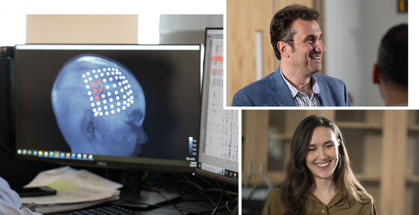
UTA Professor of Research Christos Papadelis and doctoral student Ludovica Corona set out to identify which brain cells across networks lead to epileptic episodes in children. The pair published results in the renowned journal Brain.
Using non-invasive techniques and advanced computational methods, Dr. Papadelis, Corona, and their team measured the electric and magnetic signals in the brains of 37 children and young adults with drug-resistant epilepsy who underwent neurosurgery. The patients were treated at Boston Children’s Hospital between 2011 and 2018. Through mapping, the researchers showed how seizures spread across multiple brain networks.
“For kids whose seizures cannot be controlled with medication, it is a huge burden for them and their families. Sometimes these children have multiple seizures a day,” says Papadelis, who is also the director of research for the Jane and John Justin Neurosciences Center at Cook Children’s Health Care System. “If we can disable the area where seizures originate, either with laser ablation or resective surgery, these children could become seizure-free.”
The project—a collaboration with Boston Children’s Hospital, Massachusetts General Hospital, and Harvard Medical School—was funded by the National Institute of Neurological Disorders and Stroke. It’s another example of Papadelis’ extensive work developing biomarkers for epilepsy.
Earlier this year, for example, he secured a $2.3 million grant from the same agency to explore combining two types of non-invasive imaging and brain mapping techniques to better understand seizures and epileptiform brain networks. Doing so could improve surgical precision and also open the door for children whose cases were previously considered inoperable.
“Seizures affect children throughout their lives and can have a devastating impact on development,” he says. “Our ultimate goal is to help children live free of seizures.”
Mapping the Timeline of Alzheimer’s
In a healthy brain, a web of neurons constantly fires billions of signals to other neurons and throughout the body. In the brain of an Alzheimer’s patient, however, those neurons slow, lose connections with other neurons, and eventually wither. The parts of the brain that control memory are attacked, followed by those responsible for language, reasoning, and behavior.
Alzheimer’s is devastating for patients, families, and caregivers. More than 6 million people in the United States have the disease, and by 2050 that number is expected to double. For decades, scientists have studied predictive tools to identify Alzheimer’s and its precursor, mild cognitive impairment. But these tools largely overlooked the nature of how the disease develops and progresses.
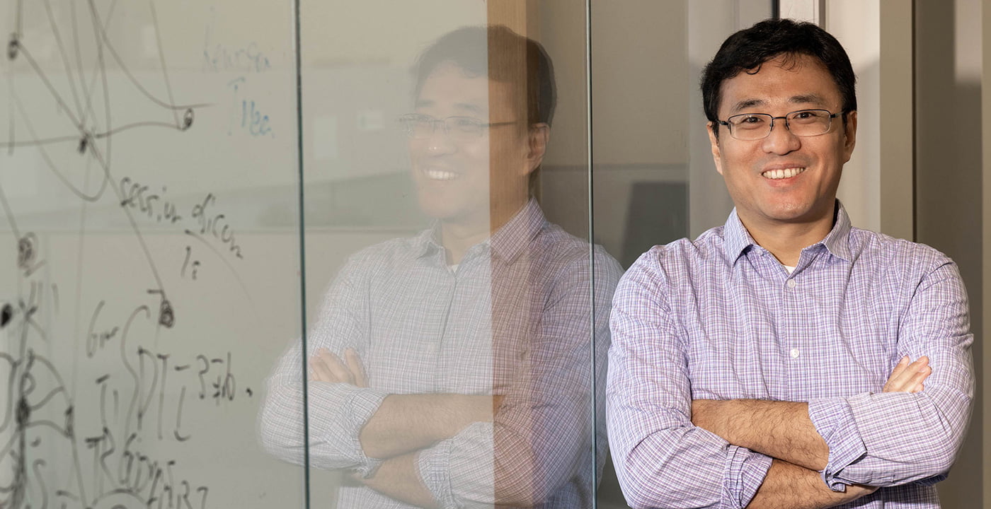
Dajiang Zhu, an associate professor of computer science and engineering, set out to develop a framework to help Alzheimer’s patients pinpoint where they fall within the disease’s progression. His goal is to enable patients and caregivers to predict the timing of its advanced stages and prepare for future care needs.
Dr. Zhu has focused his career on how machine learning and computational neuroscience can uncover solutions for pressing medical concerns. Previously, he worked with the University of North Carolina and the University of Georgia to develop deep-learning methods and tools to analyze Alzheimer’s disease and Lewy body dementia. Differentiating between the two types of dementia is challenging because of mixed pathologies and clinical symptoms.
Most recently, he and Li Wing, an associate professor of mathematics, developed a deep-learning-based framework that codes the various stages of Alzheimer’s disease in a process called “disease-embedding tree,” or DETree. Using that framework, DETree can predict how and when the disease progresses. The project is supported by more than $2 million in grants from the National Institutes of Health and the National Institute on Aging.
Zhu, director of UTA’s Medical Imaging and Neuroscientific Discovery Laboratory, described the findings in Pharmacological Research. To test the novel framework, he and his team studied the progression of the disease in 266 patients from the Alzheimer’s Disease Neuroimaging Initiative, a collaborative study launched by the National Institute on Aging in 2004. They compared results to other widely used predictive tools, then repeated the experiment numerous times using machine-learning methods. The DETree proved more accurate than other available tools.
The team believes the DETree even has the potential to predict the progression of other multi-staged neurological diseases, including Parkinson’s, Huntington’s, and Creutzfeldt-Jakob, a rare brain disorder that causes dementia.
“Individuals living with Alzheimer’s disease develop worsening symptoms at different rates,” Zhu says. “This new framework could help patients and their families better plan for the uncertainties of this complicated and devastating disease.”
You Might Also Like

Inspiration Takes Aim
Research breakthroughs can come from unexpected places. UTA professors tell us about their eureka moments.

Driving Innovation in Texas
By focusing on research important to the future economic and cultural growth of Texas, UTA is making an impact.

A Focus on Faculty
UTA has launched a $60 million faculty hiring initiative to amplify its research impact.