Life Sciences Building, Room 206
501 S. Nedderman Drive
Box 19047
Arlington, TX 76019
UTA study to focus on reducing radiation exposure from medical imaging procedures
Jin receives $500K NIH grant for project using deep learning models to analyze CT angiography scans
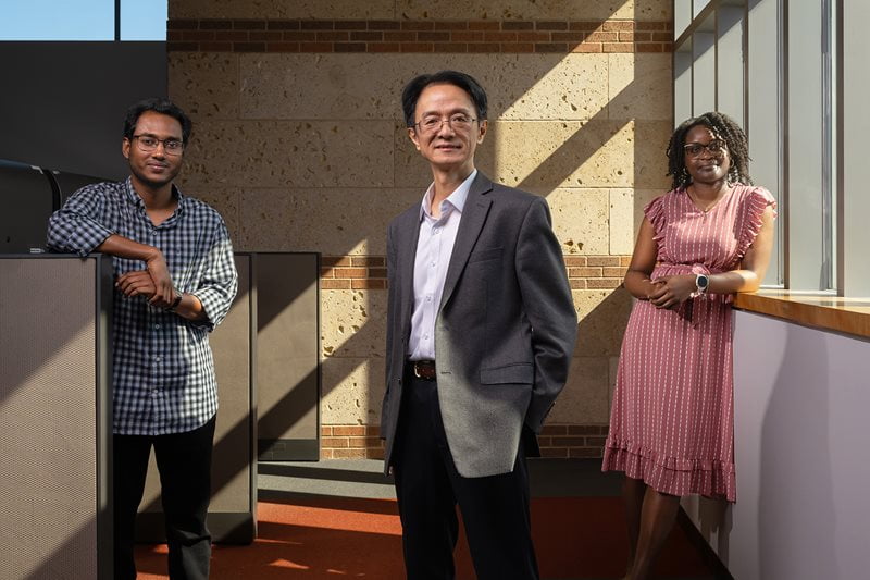
Mingwu Jin, UTA professor of physics, with graduate students Noor Rasel, left, and Darnisha Langlais, right.
A new medical physics study at The University of Texas at Arlington could substantially reduce the amount of radiation that heart patients receive during imaging procedures without sacrificing diagnostic accuracy.
Mingwu Jin, professor of physics, will lead the project, which is funded by a three-year, $579,745 grant from the National Heart, Lung, and Blood Institute, a division of the National Institutes of Health. The project is titled “Deep leaRning-bAsed reConstruction (DRAC) for Multi-Phase Cardiac CT Angiography.”
Jin’s study will focus on computed tomography angiography (CTA), an advanced imaging test that noninvasively assesses coronary artery disease, the number one killer in the world. CTA combines a CT scan with iodine contrast dye injected into a patient’s vein to create 3-dimensional pictures of blood vessels and surrounding tissues throughout the body. Doctors can use CTA to detect aneurysms, narrowed or damaged blood vessels, blood clots, tumors, and more.
One type of CTA procedure is multi-phase coronary CTA (MP-CCTA), which provides better information than single-phase CCTA and less chance of motion artifacts (blurring due to the beating heart during the procedure). However, MP-CCTA delivers a higher radiation dose, which can increase cancer risk, especially detrimental for the 80 percent of patients with negative findings.
“Lowering radiation dose in medical imaging has been vigorously pursued due to imaging prevalence and potential cancer risk of ionizing radiation,” Jin said. “Although notable progress has been made by developing advanced reconstruction methods, the dose reduction potential is far from its limit.”
One advanced reconstruction method is model-based iterative reconstruction (MBIR), which is a CT algorithm used to generate high-quality cross-sectional images from raw data. Another is 4D reconstruction, which adds time to standard 3D space, and in cardiac imaging captures clear views of fast-moving heart valves and coronary arteries without motion artifacts.
Jin said that recent breakthroughs in deep learning provide an unprecedented opportunity to greatly lower radiation dose levels through the development of 4D DRAC methods that combine MBIR with features learned from a large, high-quality MP-CCTA image database. If achieved, this could shift the conventional “individual-data-dependent” reconstruction paradigm to a revolutionary “individual-and-big-data-dependent” reconstruction paradigm.
“We hypothesize that 4D DRAC can achieve ultra-low dose for MP-CCTA, meaning around 3 millisieverts (mSv) or less, using low tube current and limited angle (fast-pitch) scan with the same or improved diagnostic accuracy,” Jin said. A millisievert is a unit used to measure radiation dose, the amount of radiation absorbed by the body.
To test the hypothesis, Jin and his team will:
1) build a synthetic patient library using computational human phantoms with real patient parameters and textures based on retrospectively collected MP-CCTA data from real patients;
2) develop image-domain DRAC (iDRAC) based on 3D and 4D deep learning and 4D DRAC methods using both CT imaging physics and features extracted by iDRAC;
3) find the minimum radiation dose that the method can achieve without compromising image quality. To evaluate the image quality, a model observer (a computer model mimicking the human observer, i.e. radiologist) will be used and validated by a small-scale human observer study.
The Mayo Clinic in Rochester, Minnesota will provide MP-CCTA patient data for the study. The project could substantially lower the radiation dose of MP-CCTA, which will have a direct impact on the standard care of stenosis detection of coronary artery disease and a broad impact to its prognostic value of monitoring high-risk plaque progression, Jin said. He added that the 4D DRAC methods could be expanded to other dynamic CT scans, where a series of CT images are acquired to see temporal changes.
“This award reflects the high quality of Dr. Jin’s research and the strength of his proposed ideas,” said Alex Weiss, professor of physics and former department chair. “I know a great deal of work goes into preparing a successful proposal of this kind. This is a significant achievement for Dr. Jin and a major benefit to our department.”
Graduate students Noor Rasel and Darnisha Langlais will work with Jin on the project, and the grant will enable Jin to hire undergraduate students in the summers and provide them with valuable research opportunities in applying AI techniques to medical imaging.
---
The UTA College of Science, a Carnegie R1 research institution, is preparing the next generation of leaders in science through innovative education and hands-on research and offers programs in Biology, Chemistry & Biochemistry, Data Science, Earth & Environmental Sciences, Health Professions, Mathematics, Physics and Psychology. To support educational and research efforts visit the giving page, or if you're a prospective student interested in beginning your #MaverickScience journey visit our future students page.