A microscope that can monitor the development of the heart
The ability to dynamically track the movement of cells is essential for modeling cellular interactions as they form organs such as the heart. But current microscope technology isn’t up to the task of capturing those movements.
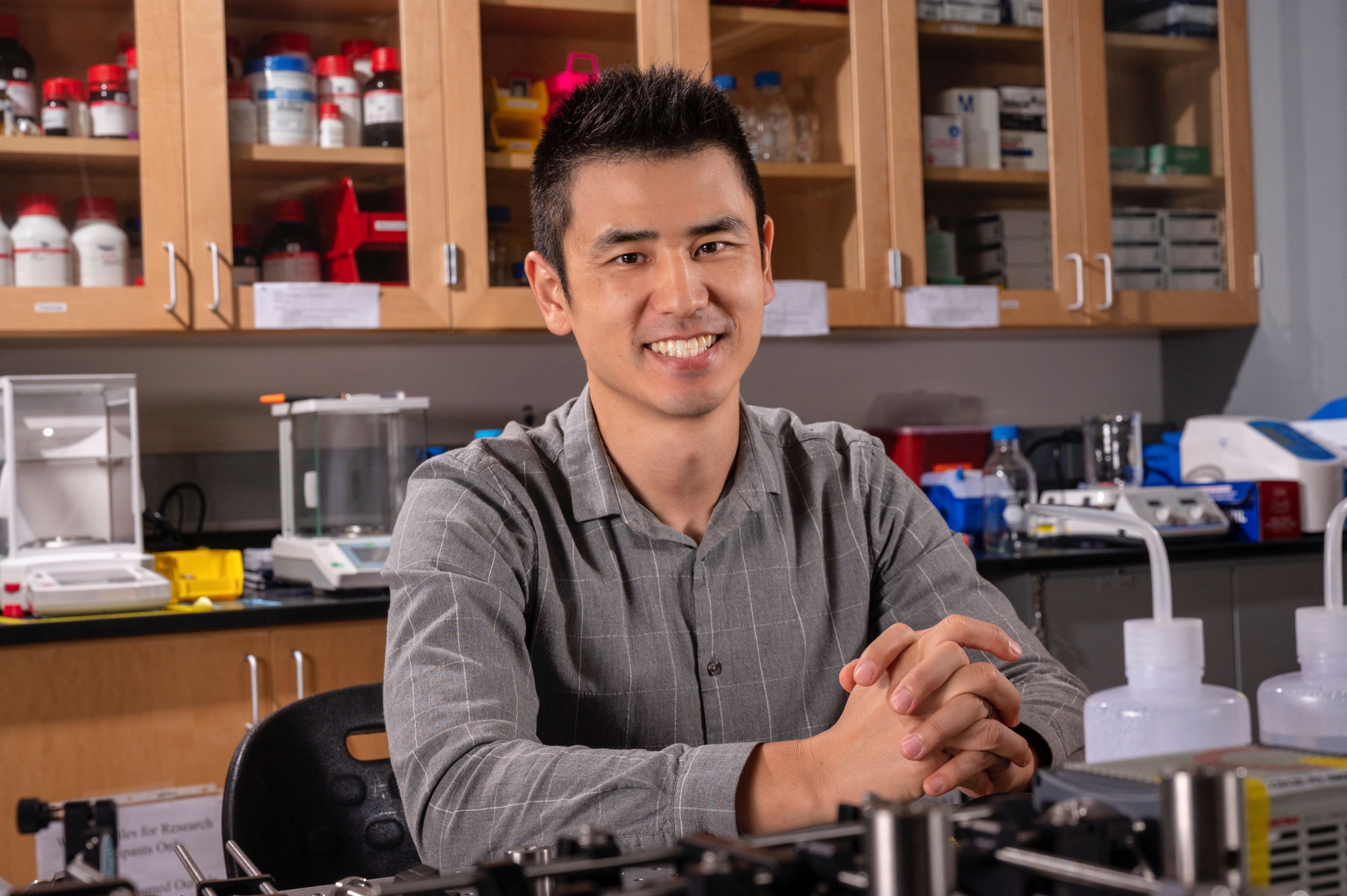
Juhyun Lee, associate professor in the Bioengineering Department at The University of Texas at Arlington, recently received a five-year, $1.94 million grant from the National Institutes of Health to develop a 4D high-resolution imaging system to quantify cell tracking.
Traditional microscopes allow users to zoom in to view an individual cell. However, doing so obscures that cell’s relationship to everything around it. Likewise, when the microscope is zoomed out to view the full field, the cell details are no longer visible.
Lee’s system will use microscopy and advanced computational post-processing to provide the next generation of imaging tools for biomedical researchers. His high-resolution microscope will zoom in and sweep left and right to scan an entire field of view. Those images will be stitched together to show a complete view, and an additional scan will create a 3D model.
“We are trying to quantify where each cell will go as the heart develops. The whole idea of this tool is to enable us to see the whole picture in high resolution,” Lee said. “Cells that form organs with dynamic motion, like the heart, are fast-moving. Using methods based on the motion and orientation of a cell, we can predict where it will go.”
Lee’s research has focused on how the heart develops, and he has received multiple previous grants focused on developing a new microscope that can capture 3D motion and add time. With this, he can construct a 4D beating heart using optical imaging techniques with fluorescent nanoparticles in a zebrafish. It is this microscope that will be used in his new grant.
Developmental biologists could eventually use Lee’s system to predict early cell development, making it valuable for detecting possible anomalies.
“The integration of technology and biology is a constant goal in the biomedical engineering field, and Dr. Lee’s work to develop imaging systems that can assist in our knowledge of cell activity and development is at the forefront of that effort,” said Michael Cho, the Alfred R. Potvin and Janet H. Potvin Endowed Chair in Bioengineering.
- Written by Jeremy Agor, College of Engineering