Life Sciences Building, Room 206
501 S. Nedderman Drive
Box 19047
Arlington, TX 76019
Jin receives $472K NIH grant for study of how to improve accuracy of stress tests
A physicist at The University of Texas at Arlington is leading a study designed to improve the accuracy of stress tests to better diagnose heart disease.
A physicist at The University of Texas at Arlington is leading a study designed to improve the accuracy of stress tests to better diagnose heart disease.
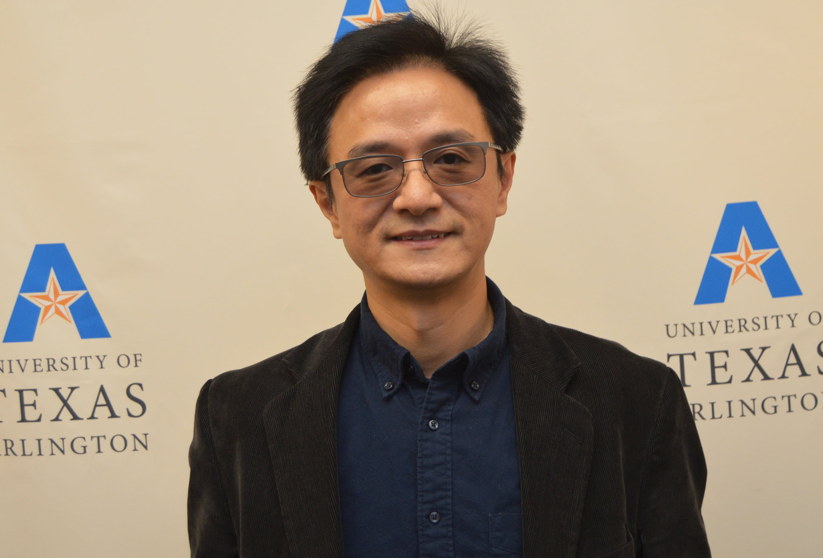
Mingwu Jin, associate professor of physics, is principal investigator of a $472,225 grant from the National Institutes of Health’s National Heart, Lung, and Blood Institute for a project titled “Attenuation Correction Strategies for Myocardial Perfusion Imaging Using Dual-Gated SPECT”.
The project’s goal is to increase the effectiveness of heart perfusion imaging – also called a nuclear stress test – which is a way of measuring how well blood flows through the heart muscle and how well the heart is pumping. The tests are very common, with as many as 8 million performed in the United States each year. The test uses small, safe doses of radioactive drugs called tracers, which mix with the blood and are taken up by the heart muscle as blood flows through the heart’s arteries.
One popular method of heart perfusion imaging is cardiac single-photon emission computed tomography (SPECT) imaging. One of the drawbacks to traditional SPECT imaging is that it normally takes more than 10 minutes and eliminating all patient motion during the test is essentially impossible. Movement can cause blurring of the images and make diagnoses more difficult. Another problem is attenuation artifacts, which are caused by a reduction in the strength of the imaging signal when it travels through body tissues of different densities, such as breast tissue, the chest wall, and organs under the diaphragm.
“In order to minimize the motion artifacts, the SPECT data acquisition can be synchronized with the heartbeat and the breath rhythm, which is called dual-gated SPECT,” Jin said. “With sophisticated physics modeling in image reconstruction algorithms and cone-beam CT (CBCT) data, we will develop and investigate strategies to correct photon attenuation artifacts of dual-gated SPECT.”
The research is highly relevant to public health because the development of respiratory motion matched attenuation correction using low-dose X-ray CBCT for myocardial perfusion imaging using SPECT could substantially reduce image artifacts and improve diagnostic accuracy for heart disease.
“The proposed method, which holds the potential to eliminate these attenuation artifacts using advanced image reconstruction and processing techniques, will be evaluated cost-effectively by mass computer simulations and a preliminary patient study,” Jin said.
The project is a collaborative effort between UTA, the University of Massachusetts Medical Center (UMMC), and the University of Texas Southwestern Medical Center (UTSW) in Dallas. Jin explained that UTA will lead the effort involving study design, algorithm development and data analysis. UMMC collected SPECT/CBCT data for more than 1,500 patients, and will provide real patient data and clinical inputs. UTSW will provide access to the CBCT scanner and help on algorithm development.
The project will also provide opportunities for UTA students to be professionally trained and to gain valuable experience in medical imaging research with clinical collaborators. Shiwei Zhou and Akolade Adebayo, Ph.D. students in Jin’s lab, are working on the project, and some undergraduate students will also have the opportunity to join in the study, Jin said.
Jin and his colleagues hope that the project will help provide solutions to two important issues in medical imaging.
“First, the project will answer the question of which type of heart perfusion defects will benefit most from this sophisticated SPECT imaging protocol,” he said. “Secondly, if the project is successful, a novel attenuation correction method can be applied to dual-gated SPECT, which will lead to the artifact-free perfusion images for more accurate, reproducible and faster diagnosis of heart diseases.”
Since 2016, Jin has been awarded more than $1.1 million in funding for projects aimed at improving medical imaging technology.
Alex Weiss, professor and chair of the UTA Department of Physics, said that Jin’s latest study could lead to advances in research focusing on health and the human condition as well as data driven discovery, which are two of the main points of emphasis of UTA’s Strategic Plan 2020.
“Dr. Jin is addressing important issues in medical imaging and his work will help to improve the technology utilized in the diagnosis and treatment of millions of patients,” Weiss said. “His research is representative of the outstanding contributions being made by our innovative faculty in the Department of Physics.”
Jin received a B.S. degree in Space Physics and an M.E. degree in Communication and Information Systems from Peking University, Beijing, China, in 1997 and 2001, respectively. He earned a Ph.D. in Electrical Engineering from Illinois Institute of Technology in Chicago in 2007. He worked as a research associate and an instructor in radiology at the University of Colorado at Denver from 2007-10, and came to UTA as a research assistant professor in 2011. He became an assistant professor of physics in 2014 and was promoted to associate professor in 2017.