634 Nedderman Hall
Box 19019
416 Yates Street
Arlington, TX 76019-0019
Healthcare
Biomedical Imaging, Bioinstrumentation, Regenerative Medicine and Tissue Engineering, Omics
Recent Highlights

Center for Assistive Technologies to Enhance Human Performance - iPerform
The iPerform Center is an NSF-funded Industry-University Cooperative Research Center (I/UCRC) bringing professors and scientists at the University of Texas at Arlington and the University of Texas at Dallas together to advance basic and applied research in Assistive Technologies to enhance human performance.
Resources
Cancer and Regenerative Medicine Laboratory
The lab has complete cell culture, bacteria culture, histology, surgical equipment, and imaging equipment sufficient to carry out a wide variety of preclinical research. This lab provides essential expertise and the facility to translate scientific discoveries into clinical practice to benefit humanity.
Cardiovascular Bioengineering Lab
Voxel super-resolution light-sheet microscopy to image centimeter to micron-scale specimens. AI-driven 4D imaging system to capture the rapid dynamic motions of biological sample. Computational tool to quantify biomechanical cues to understand cardiac morphogenesis. Extensive biomedical research equipment to mimic the environment of human body.
Biomolecular Imaging Lab
Multi-modality imaging capabilities to probe mechanobiological interactions over multiple length and time scales, including atomic force microscopy, two-photon microscopy, and nanovid microscopy, in vitro explosive blast system to simulate shock wave exposure on the battlefield.
Nanomedicine Lab
Temperature-controlled ultracentrifuge, hyperspectral microscopy to characterize micro- and nano-scale materials, freeze dry system to lyophilize biological samples, nanoparticle characterization capability to determine the particle size and surface charges, exceptionally sensitive spectrophotometer to utilize wavelengths from UV to near infrared spectrum.
Multi-Interprofessional Center for Health Informatics (MICHI)
MICHI draws from strengths and knowledge of multiple professions and disciplines with the ultimate goal of transitioning from “sick care” to “health care.” Through strategic partnerships, the Center uses health informatics approaches to support data-driven initiatives that impact health and introduce and assess various enabling tools into all aspects of living and learning.
<< Back to Research
Areas
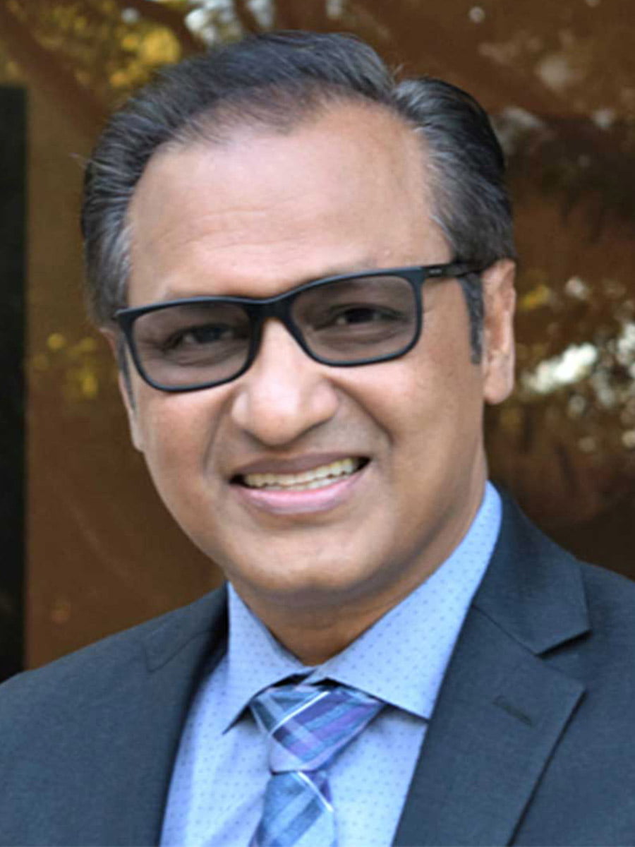
Ashfaq Adnan, Ph.D.
Mechanical and Aerospace Engineering
George Alexandrakis, Ph.D.
Bioengineering
Marion Ball, Ph.D.
Bioengineering

Khosrow Behbehani, Ph.D.
Bioengineering
Alan Bowling, Ph.D.
Mechanical and Aerospace Engineering
Ye Cao, Ph.D.
Materials Science and Engineering

Michael Cho, Ph.D.
Bioengineering
Gautam Das, Ph.D.
Computer Science and Engineering
Digant Davé, Ph.D.
Bioengineering
Yaowu Hao, Ph.D.
Materials Science and Engineering

Haiying Huang, Ph.D.
Mechanical and Aerospace Engineering

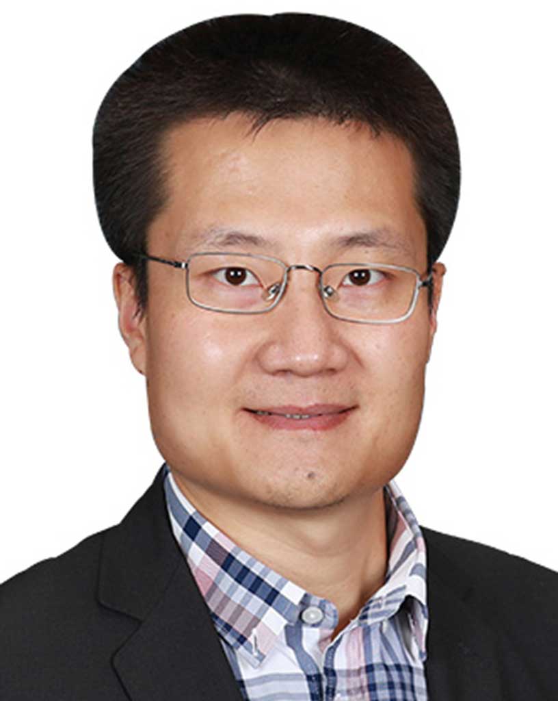
Junzhou Huang, Ph.D.
Computer Science and Engineering
Yi Hong, Ph.D.
Bioengineering
Young-Tae Kim, Ph.D.
Bioengineering
Seong-Jin Koh, Ph.D.
Materials Science and Engineering
Juhyun Lee, Ph.D.
Bioengineering
Jun Liao, Ph.D.
Bioengineering
Hanli Liu, Ph.D.
Bioengineering

Jacob Luber, Ph.D.
Computer Science and Engineering

Fillia Makedon, Ph.D.
Computer Science and Engineering
Alice Sun, Ph.D.
Electrical Engineering

Liping Tang, Ph.D.
Bioengineering

Baohong Yuan, Ph.D.
Bioengineering
Kyungsuk Yum, Ph.D.
Materials Science and Engineering
Weidong Zhou, Ph.D.
Electrical Engineering
Dajiang Zhu, Ph.D.
Computer Science and Engineering
Related Faculty
Ashfaq Adnan, Ph.D.
Mechanical and Aerospace Engineering
George Alexandrakis, Ph.D.
Bioengineering
Marion Ball, Ph.D.
Bioengineering
Khosrow Behbehani, Ph.D.
Bioengineering
Alan Bowling, Ph.D.
Mechanical and Aerospace Engineering
Ye Cao, Ph.D.
Materials Science and Engineering
Michael Cho, Ph.D.
Bioengineering
Gautam Das, Ph.D.
Computer Science and Engineering
Digant Davé, Ph.D.
Bioengineering
Yaowu Hao, Ph.D.
Materials Science and Engineering
Haiying Huang, Ph.D.
Mechanical and Aerospace Engineering
Junzhou Huang, Ph.D.
Computer Science and Engineering
Yi Hong, Ph.D.
Bioengineering
Young-Tae Kim, Ph.D.
Bioengineering
Seong-Jin Koh, Ph.D.
Materials Science and Engineering
Juhyun Lee, Ph.D.
Bioengineering
Jun Liao, Ph.D.
Bioengineering
Hanli Liu, Ph.D.
Bioengineering
Jacob Luber, Ph.D.
Computer Science and Engineering
Fillia Makedon, Ph.D.
Computer Science and Engineering
Alice Sun, Ph.D.
Electrical Engineering
Liping Tang, Ph.D.
Bioengineering
Baohong Yuan, Ph.D.
Bioengineering
Kyungsuk Yum, Ph.D.
Materials Science and Engineering
Weidong Zhou, Ph.D.
Electrical Engineering
Dajiang Zhu, Ph.D.
Computer Science and Engineering